| [1] |
Wilmut I, Schnieke A E, McWhir J, et al. Viable offspring derived from fetal and adult mammalian cells[J]. Nature, 1997,385(6619):810-813.
URL
pmid: 9039911
|
| [2] |
Baguisi A, Behboodi E, Melican A D, et al. Production of goats by somatic cell nuclear transfer[J]. Nature Biotech, 1999,17(5):456-461.
doi: 10.1038/8632
URL
|
| [3] |
Cibelli J B, Stice S L, Golueke P J, et al. Cloned transgenic calves produced from nonquiescent fetalfibroblasts[J]. Science, 1998,280(5367):1256-1258.
URL
pmid: 9596577
|
| [4] |
Betthauser J, Forsberg E, Augenstein M, et al. Production of cloned pigs from in vitro systems[J]. Nature Biotech, 2000,18(10):1055-1059.
|
| [5] |
Wakayama T, Yanagimachi R. Cloning of male mice from adult tail-tip cells.Nat[J]. Genet, 1999,22(2):127-128.
|
| [6] |
Galli C, Lagutina I, Crotti G, et al. Pregnancy:a cloned horse born to its dam twin[J]. Nature, 2003,424(6949):635.
doi: 10.1038/424635a
URL
pmid: 12904778
|
| [7] |
Wani N A, Wernery U, Hassan F A, et al. Production of the first cloned camel by somatic cell nuclear transfer[J]. Biology Of Reproduction, 2010,82(2):373-379.
doi: 10.1095/biolreprod.109.081083
URL
pmid: 19812298
|
| [8] |
Debra K, Berg, Li C Y, et al. Red Deer Cloned from Antler Stem Cells and Their Differentiated Progeny[J]. Biology Of Reproduction, 2007,77(3):384-394.
URL
pmid: 17522075
|
| [9] |
Cibelli J B, Campbell K H, Seidel G E, et al. The health profile of cloned animals[J]. Nat Biotechnol, 2002,20(1):13-14.
doi: 10.1038/nbt0102-13
URL
pmid: 11753346
|
| [10] |
Constant F, Guillomot M, Heyman Y, et al. Large offspring or large placenta syndrome Morphometric analysis of late ges-tation bovine placentomes from somatic nuclear transfer pregnancies complicated by hydrallantois[J]. Biol Reprod, 2006,75(1):122-130.
doi: 10.1095/biolreprod.106.051581
URL
pmid: 16571872
|
| [11] |
Chavatte-Palmer P, Heyman Y, Richard C, et al. Clini-cal, hormonal, and hematologic characteristics of bovine calves derived from nuclei from somatic cells[J]. Biol Reprod, 2002,66(6):1596-1603.
doi: 10.1095/biolreprod66.6.1596
URL
pmid: 12021036
|
| [12] |
Miglino M A, Pereira F T, Visintin J A, et al. Placentation in cloned cattle: Structure and microvascular architecture[J]. Theriogenology, 2007,68(4):604-617.
URL
pmid: 17568663
|
| [13] |
郭磊, 李慧, 韩之明. DNA甲基化和组蛋白修饰在克隆动物发育过程中的作用[J]. 遗传, 2010,32(8):762-768.
|
| [14] |
杨荣荣, 李相运. DNA甲基化与克隆动物的发育异常[J]. 遗传, 2007,29(9):1043-1048.
|
| [15] |
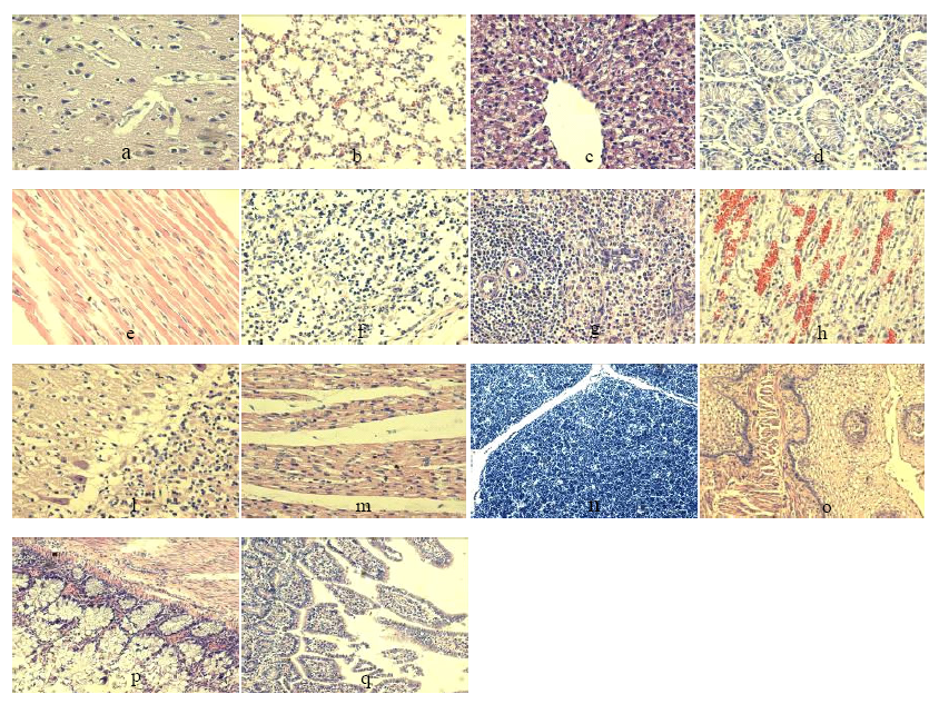
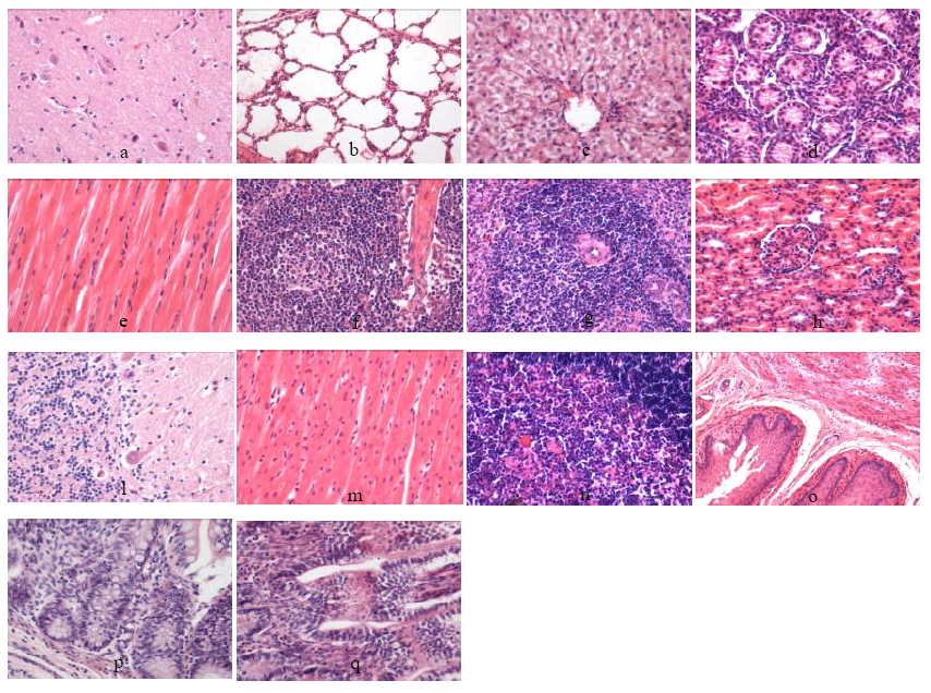
顾玉芳, 李善姬, 吴素清. 内蒙古首例体细胞克隆牛2日龄死亡后的病理学观察[J]. 长江大学学报, 2007,4(4):30-32.
|
| [16] |
王晓丽, 蒋建荣, 徐莉萍, 等. 一例体细胞克隆水牛肺、脾脏的组织结构观察[J]. 湖北农业科学, 2010,49(6):1417-1418.
|
| [17] |
袁苏娅, 姬丽娜, 王勇胜, 等. 1例转基因体细胞克隆牛主要脏器的组织病理学观察[J]. 中国兽医学报, 2013,23(1):113-118.
|
| [18] |
王晓丽, 韦精卫, 房慧伶, 等. 一例体细胞克隆黄牛肺脏的组织结构观察[J]. 中国畜牧兽医, 2009,36(2):138-140.
|
| [19] |
Agabiti Rosei E, Muiesan M L. Prognostic significance of left ventricular hypertrophy regression[J]. Adv Exp Med Biol, 1997,432:199-205.
doi: 10.1007/978-1-4615-5385-4_22
URL
pmid: 9433527
|
| [20] |
张荣华, 王曦, 李武峰. 哺乳动物克隆技术研究进展[J]. 山西农业科学, 2017,45(9):1577-1582.
|
| [21] |
Leal Yepes F A, Mann S, Overton T R, et al. Effect of rumen-protected branched-chain amino acid supplementation on production- and energy-related metabolites during the first 35 days in milk in Holstein dairy cows[J]. Journal of Dairy Science, 2019,10:5657-5672.
|
| [22] |
Zhao K X, Jin X, Ji J T, et al. Individual identification of Holstein dairy cows based on detecting and matching feature points in body images[J]. Biosystems Engineering, 2019,10:128-139.
|
| [23] |
Piantoni P, Lock A L, Allen M S. Corrigendum to “Saturated fat supplementation interacts with dietary forage NDF content during the immediate postpartum period in Holstein cows: Energy balance and metabolism”[J]. Journal of Dairy Science, 2019,98(3):3323-3334.
|
| [24] |
Piantoni P, Lock A L, Allen M S. Saturated fat supplementation interacts with dietary forage neutral detergent fiber content during the immediate postpartum and carryover periods in Holstein cows: Production responses and digestibility of nutrients[J]. Journal of Dairy Science, 2015,5:3309-3322.
|
| [25] |
Khosravi M, Rouzbehan Y, Rezaei M, et al. Total replacement of corn silage with sorghum silage improves milk fatty acid profile and antioxidant capacity of Holstein dairy cows[J]. Journal of Dairy Science, 2018,10:10953-10961.
|
 )
)