| [1] |
成仿云, 李嘉珏, 陈德忠. 中国野生牡丹自然繁殖特性研究[J]. 园艺学报, 1997, 24(2):180-184.
|
| [2] |
李嘉珏, 陈德忠, 于玲, 等. 大花黄牡丹分类学地位的研究[J]. 植物研究, 1998, 18(2):152-155.
|
| [3] |
王文华. 西藏大花黄牡丹资源保护与利用[J]. 现代农业科技, 2016, 13:188,193.
|
| [4] |
权红, 兰小中. 濒危植物大花黄牡丹光合特性及日进程研究[J]. 种子, 2013, 32(5):29-32,38.
|
| [5] |
李建平. 西藏大花黄牡丹抗絮状表皮癣菌和断发毛癣菌活性物质研究[D]. 哈尔滨: 黑龙江大学, 2015.
|
| [6] |
杨小林, 罗健, 鲍隆友. 濒危植物大花黄牡丹种群结构与分布格局[J]. 西南林学院学报, 2006, 26(6):6-9.
|
| [7] |
WU S H, WU D G, CHEN Y W. Chemical constituents and bioactivities of plants from the genus Paeonia[J]. Chemistry & biodiversity, 2010, 7(1):90-104.
|
| [8] |
张晓骁. 中国野生牡丹种质资源调查评价研究[D]. 杨凌: 西北农林科技大学, 2018.
|
| [9] |
邢震, 张启翔, 次仁. 西藏大花黄牡丹生境概况初步调查[J]. 江苏农业科学, 2007, 35(4):250-254.
|
| [10] |
郑雨, 赵玉文, 费文群, 等. 干旱胁迫下西藏2种牡丹幼苗叶绿素含量和光合特征比较[J]. 高原农业, 2018, 2(5):453-461,557.
|
| [11] |
贾文庆, 王艳丽, 郭英姿, 等. 大花黄牡丹花粉萌发及贮存特性[J]. 林业科学, 2021, 57(2):82-92.
|
| [12] |
陈庭巧, 袁涛, 乔红雍, 等. 大花黄牡丹二次枝对结实率的影响及胚珠败育生理机制研究[J]. 西北农林科技大学学报(自然科学版), 2022, 50(9):39-52.
|
| [13] |
陈庭巧, 袁涛, 解梦雨, 等. 引种栽培条件下大花黄牡丹二次枝及顶芽发育的初步研究[J]. 北京林业大学学报, 2022, 44(6):106-114.
|
| [14] |
CHEN T Q, SUN Y, YUAN T. Transcriptome sequencing and gene expression analysis revealed early ovule abortion of Paeonia ludlowii[J]. BMC Genomics, 2023, 24(1):78.
|
| [15] |
张超奇. 大花黄牡丹籽油精炼过程中化学成分的变化及其功能成分研究[D]. 拉萨: 西藏大学, 2021.
|
| [16] |
徐慧, 姚霞珍, 佟珂珂, 等. 3种牡丹花器官不同部位挥发性成分分析[J]. 南京林业大学学报(自然科学版), 2023, 47(3):63-69.
doi: 10.12302/j.issn.1000-2006.202203039
|
| [17] |
曾秀丽, 王亮生, 潘光堂, 等. 西藏野生黄牡丹和大花黄牡丹的色素分析[J]. 园艺学报, 2012, 39(增刊):2744.
|
| [18] |
李杰, 旦真次仁, 许晓嘉, 等. 西藏大花黄牡丹花朵氨基酸组成和矿质元素比较分析[J]. 云南农业大学学报(自然科学), 2017, 32(6):1058-1063.
|
| [19] |
ADKI K M, KULKARNI Y A. Chemistry, pharmacokinetics, pharmacology and recent novel drug delivery systems of paeonol[J]. Life sciences, 2020, 250:117544.
|
| [20] |
肖超妮, 王培, 马翠霞, 等. 牡丹不同根部位的代谢物分布[J]. 波谱学杂志, 2015, 32(4):648-660.
doi: 10.11938/cjmr20150410
|
| [21] |
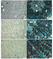
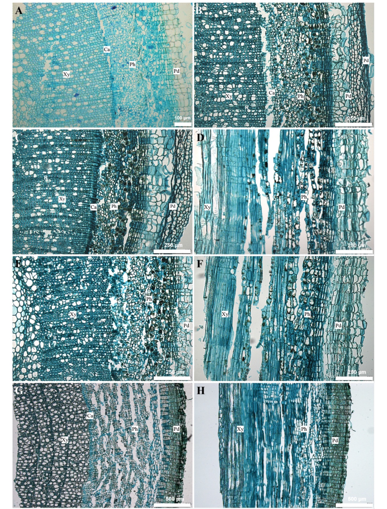
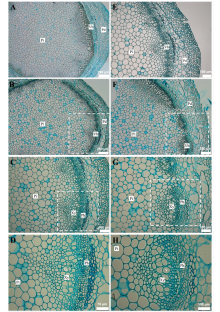
张华, 贾军涛. 大花黄牡丹营养器官形态解剖学观察[J]. 高原农业, 2018, 2(1):57-60.
|
| [22] |
杨虎彪, 李晓霞, 罗丽娟. 植物石蜡制片中透明和脱蜡技术的改良[J]. 植物学报, 2009, 44(2):230-235.
|
| [23] |
王旭初, 史敏晶, 陈月异, 等. 无患子科树木细子龙营养贮藏蛋白质的分离鉴定[J]. 热带作物学报, 2004, 25(4):78-83.
|
| [24] |
成妍, 乔宁, 梁燕平, 等. 辣椒小孢子胚胎发生早期的组织化学染色[J]. 黑龙江农业科学, 2019(8):18-21.
|
| [25] |
魏芳, 郑乾坤, 罗世巧, 等. 橡胶树树皮和木质部淀粉和可溶性糖含量测定[J]. 热带农业科学, 2014, 34(4):9-13.
|
| [26] |
TIAN W M, WU J L, HAO B Z, et al. Vegetative storage proteins in the tropical tree Swietenia macrophylla: Seasonal fluctuation in relation to a fundamental role in the regulation of tree growth[J]. Canadian journal of botany, 2003, 81(5):492-500.
|
| [27] |
朱艳梅, 罗兴录, 颜国彪, 等. 木薯蔗糖合成、转运与块根淀粉积累关系研究[J]. 广西植物, 2016, 36(12):1492-1497.
|
| [28] |
张秀玲. 如何观察植物细胞的后含物和质体[J]. 生物学通报, 2006, 41(4):53-54.
|
| [29] |
CHEN J, SHEN Y Q, YAO Y J, et al. Research progress of superabsorbent polymer materials[J]. Chemical industry and engineering progress, 2022, 41(11):5925-5935.
doi: 10.16085/j.issn.1000-6613.2022-0085
|
| [30] |
由继红, 董春光, 史晓昆. 小麦叶片可溶性糖含量测定方法的研究[J]. 实验室科学, 2021, 21(2):27-29.
|
| [31] |
赵溪竹, 杨洪, 郭冰冰, 等. 源库关系研究现状及在橡胶树排胶机理研究中的展望[J]. 热带作物学报, 2023, 44(12):2506-2513.
doi: 10.3969/j.issn.1000-2561.2023.12.016
|
 ), 张世鑫2, 蒋毅1,2, 郑硕恒1,2, 戴明鋆1,2, 史敏晶2, 田维敏2,3, 张华1(